By Digital Smile Design
⋅ 7 min read
⋅ Updated Jul, 2025
Ever wondered why your intraoral scans aren't quite delivering the perfect results you envisioned? In a series of compelling Instagram videos leading up to the Intraoral Scanner Festival earlier this year, Christian Coachman peeled back the curtain on the often-overlooked pitfalls of everyday intraoral scanning.
From tricky bite registrations to complex full-arch implant cases, he meticulously dissected real-world challenges, revealing exactly where things can go awry and, crucially, what you can do to elevate your outcomes. His message resonated: owning a scanner is merely the first step; true mastery is where the magic happens. Dive into his insights below to unlock the precise know-how you need to conquer these hidden problems and truly master your intraoral scanning.
Top intraoral scanning tips from Christian Coachman
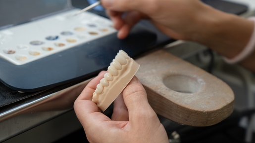
1. Poor scanning is a very expensive mistake
Christian points out that inaccurate scanning can lead to major consequences such as wasted appointments and costly remakes, and even long-term treatment failures. While many dentists assume that using a scanner automatically ensures accuracy, Christian warns that scanners are not magic solutions. Most dentists are still making mistakes without realizing it and the only way to improve is by mastering scanning as a true clinical skill.
Don’t assume your scanner guarantees accuracy: Many scans contain distortions and issues that go unnoticed.
Understand the cost of mistakes: Wrong data leads to wasted time, remakes and procedures that may fail too early.
Recognize that scanning is a technique: Mastering it requires more than just owning the equipment.
Push beyond the basics: Dental companies may teach the fundamentals but there is much more to learn from experienced users and evidence-based sources.
Question your expertise: Even dentists who scan daily may still have recurring issues that only become obvious when seen at scale in the lab.
Learn from lab feedback: Labs see high volumes of cases and repeated patterns, offering valuable insight into what is going wrong.
Involve your whole team: Assistants often perform the scans, so they need proper training to avoid introducing errors into the workflow.
2. Bite registration
Christian highlights a critical issue in digital dentistry: scanning the bite. Although it may look simple in presentations, bite registration is often done poorly – even by experienced clinicians. Digital tools can create distortions and the assumption that scanners are always precise is a major pitfall. The process requires thoughtful clinical decisions and careful technique, both at the chair and in the lab. Christian emphasizes that improving your scanning technique, starting with the bite, is one of the best investments you can make.
Recognize that digital is not magic: Scanners can create distortions when capturing the bite. You need to know how to avoid or reduce these issues clinically and digitally.
Decide on the correct bite before scanning: Evaluate whether to maintain the patient's current bite or find a better one. If a new bite is needed, learn how to deprogram the old one first.
Understand the difference between jaw positions: Know if you are registering maximum intercuspation or centric relationship and why that matters.
Master clinical and lab strategies: There are many techniques and tools, both in the clinic and in the lab, that help achieve an accurate bite registration.
Support your lab's role: Labs should analyze the scanned bite, question whether it looks ideal, and use software tools to improve the registration if needed.
Reduce complications during restoration: The more precise the bite scan, the fewer adjustments will be needed in the mouth later on.
Make scanning technique a priority: Improving how you scan, starting with bite registration, is more important than buying new tools or technology.
3. Jaw motion
Christian discusses the importance of recording a patient's real jaw movement. While articulators can simulate motion, they do not capture the full picture, especially in complex functional cases. He explains why this type of technology is becoming essential in modern dentistry and how it can influence diagnosis, treatment planning and the design of restorations. He also raises key questions every restorative dentist should consider before investing in jaw tracking systems.
Understand the limitations of articulators: Both analog and digital articulators are not sufficient for more complex occlusion and functional cases.
Recognize the value of real jaw motion: Recording the true movement of the mandible is an important step in understanding TMJ function, diagnosing problems, and planning the right treatment.
Use jaw motion data to improve restorations: Sending real jaw movement, including chewing, to the lab helps design restorations that better fit functional needs.
See it as part of the patient avatar: Jaw motion is one of the five key components used to create a complete digital representation of the patient.
Think ahead: Christian believes recording real jaw motion is not just helpful, but part of the future of dentistry.
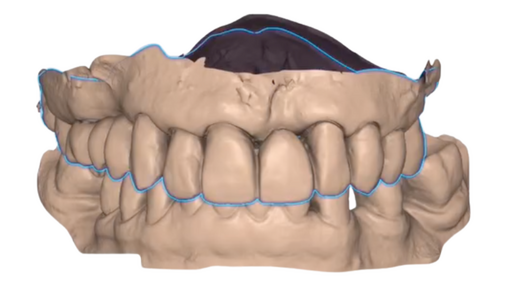
4. Occlusal artificial collisions and occlusal artificial gaps
Two common but serious problems that occur during bite scanning are occlusal artificial collisions and occlusal artificial gaps. These issues happen when the upper and lower scans are not aligned realistically, leading to inaccurate restorations. The result is more time-consuming adjustments in the mouth and compromised outcomes. Many dentists are unaware of these scan errors, making it essential to understand what to look for and how to avoid them.
Check for occlusal artificial collisions: This happens when the upper scan goes inside the lower scan, creating an unrealistic overlap.
Look out for occlusal artificial gaps: These occur when the upper and lower scans are not touching in areas where real contact exists.
Understand the impact on lab work: When these scan errors occur, the lab designs restorations based on incorrect data, which leads to poor fit and increased chairside adjustments.
Learn the scan evaluation techniques: Understanding how to identify and fix these issues early in the scanning process can save time and improve clinical results.
5. STL and CBCT overlapping
This video covers the consequences of mismatches between intraoral scans and CBCT files. Even a small misalignment, such as one millimeter, can affect multiple parts of the implant workflow. These mismatches are more common than most dentists realize and can compromise implant planning, surgical guide design, surgical execution, and the fit of the final prosthesis. Christian emphasizes the importance of learning how to detect and correct this issue.
- Be aware of STL and CBCT mismatches: Even small mismatches between these files can have a big impact on treatment outcomes.
- Understand the risk with full arch cases: In multiple implant or full arch cases, mismatches often lead to bridges that do not fit passively.
6. Scan bodies
A common but underestimated problem in implant dentistry is the distortion that occurs when scanning implant scan bodies. Although it may seem like a simple and predictable process, many dentists still make small mistakes that lead to inaccurate files. These errors can compromise restorations, even in straightforward cases like single crowns or small bridges, and disrupt the digital workflow.
Do not assume implant scan body scans are always accurate: Small distortions are more common than expected and can affect both single and multi-unit restorations.
Understand the consequences of inaccuracy: A distorted scan body file will lead to an incorrect restoration and create issues during fitting.
Learn the techniques to avoid mistakes: Every dentist must know the key scanning tips that prevent distortion and maintain precision in implant cases.
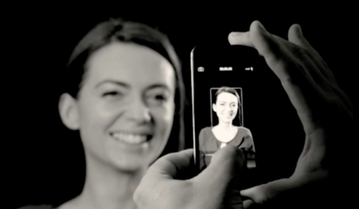
7. Building the patient avatar
The patient avatar is a complete 3D digital representation of the patient’s oral and facial structures. To build this avatar, five core components are needed: intraoral static, intraoral motion, facial static, facial motion, and inner tissues. Christian explains how each type of data is captured, the tools available and the challenges that come with combining them accurately. If the files are mismatched, it can negatively affect diagnostics, treatment planning and the final outcome.
Know the five components of the patient avatar: Intraoral static (scans), intraoral motion (jaw tracking or digital articulator), facial static (photos or scans), facial motion (video), and inner tissues (CBCT).
Capture and share complete data: Each component needs to be collected with enough quality to support planning and design, and shared with the lab for device and restoration manufacturing.
Match files correctly in software: To align files properly, you need common surface areas between them and the right tools and techniques to perform the match.
Understand the tools available: Learn which technologies and software options can help with both capturing and matching the different data types accurately.
Follow a step-by-step approach: Capture high-quality information, ensure overlaps have enough shared surface, and use the correct workflow for precise digital integration.
8. Mesh crash
A frequent issue that affects scan accuracy but often goes unnoticed by dentists? The mesh crash. A mesh crash happens when parts of a scan are missing or incomplete, creating holes that the scanner software tries to fill in. This leads to misadaptation and, when it involves critical areas like the prep surface or margin, the result can be a poorly fitting restoration that could have been avoided with better scanning and quality control.
Do not rely on automatic fixes: When the scanner fills in missing areas, it cannot recreate the true topography of the tooth, especially around prep margins.
Understand the impact on restoration fit: Mesh crashes lead to misfits, especially if they affect the prep surface or margin, which are often the hardest areas to scan.
Learn how to avoid mesh crashes: There are scanning techniques that help reduce the chance of creating these issues in the first place.
Check and fix your scans before sending: Dentists must evaluate the scan quality, identify any mesh crashes, and fix them instead of passing the problem to the lab.
9. Scanning MIP (Maximum Intercuspation)
Bite Finder is a digital tool designed to improve how dentists register and transfer maximum intercuspation. Christian explains why accurate bite registration is critical for diagnostics, treatment planning and restoration design, and how common issues in digital bite scans can create unrealistic contacts or gaps. Bite Finder offers a simple way to bring upper and lower arches together based on real contact points, similar to how technicians used to do it by hand with stone models.
Know the importance of maximum intercuspation: Whether or not the case will be restored in this position, registering and understanding this jaw relationship is essential for diagnosis and planning.
Be aware of common bite scan problems: Issues like artificial gaps or collisions in the scan can distort the occlusal surface and lead to faulty restorations.
Use software to evaluate the bite: Always check the scan in your software, look at the occlusal gram, and watch for unrealistic areas that could affect the result.
Be proactive with suspicious scans: If something does not look right, compare with the mouth or models, and use tools to find a better intercuspation before proceeding.
Look for simple solutions: Tools like Bite Finder are fast and easy to use, and can greatly reduce errors in bite registration.
10. Scanning full-arch implants
Scanning full-implant arches remains one of the most complex challenges in digital dentistry. Christian breaks down the technical limitations of intraoral scanners, including how micro stitching distortions accumulate as the scan progresses around the arch. These distortions often go unnoticed in tooth-supported restorations, but for implant-supported prostheses, they can prevent a passive fit. Christian outlines various technologies and strategies that are now being used to reduce or eliminate these distortions, highlighting the importance of understanding each option and its cost-benefit.
Understand the scanning limitation: Intraoral scanners work by stitching many small scans together, which introduces micro distortions, especially around the curve of a full arch.
Know the difference in case types: Small distortions may not affect tooth-supported crowns, but they can cause serious fit problems in full-arch implant cases.
Be aware of new technologies: Some scanners now claim to handle full-arch cases with better accuracy.
Explore photogrammetry: This technology allows accurate capture of implant positions and has evolved to include lower-cost solutions and even smartphone applications.
Match the tool to your volume: The best solution depends on how many full-arch cases you handle. Higher volume may justify the investment in more advanced technology.
Consider business models: Some labs may offer or rent technologies to dentists to improve workflow precision and ensure better outcomes.
11. Scanning gingival information
Christian focuses on an often-overlooked part of scanning: capturing gingival information. While most discussions around scanning technique concentrate on teeth, models and occlusion, Christian highlights the critical role of soft tissue data. Properly scanning the gingiva, especially around the submergence and emergence profiles, can dramatically improve the final outcome of a restoration. He explains how this information can be captured, analyzed and shared with the lab to influence both esthetics and function.
Do not ignore the gingiva: The interface between the restoration and the soft tissue plays a major role in how the final result looks and functions.
Adopt simple scanning techniques: There are easy and practical methods to record gingival information accurately, and better analyse the information.
Communicate with the lab: Send complete and clear gingival data so the lab can design restorations that respect not just the prep and occlusion but also the gingiva.
12. A real problem in restorative dentistry
Christian addresses the widespread quality issues in impressions, challenging dentists to have honest conversations with their labs about the percentage of impressions and scans that are actually usable.
Ask your lab for honest feedback: Find out what percentage of your scans or impressions are not good enough and be open to the answer.
Understand the reality: The percentage of low-quality impressions and scans - especially for tooth preparations and implants - is higher than most dentists think.
Make quality your goal: Aim to be among the small group of dentists who consistently send high-quality scans to the lab.
Start with a high-end scan: This is the foundation of the restorative workflow and critical for successful smile rehabilitation and prosthetic results.
Learn and apply the right techniques: Use the available tips, tricks and strategies to upgrade your scanning approach and deliver better data to your lab.
What every lab wants to see
What do labs consider the gold standard when it comes to scan quality? Christian talks about a perfect case in which the scan shows excellent overlap between the prep and provisional scans, clean and clearly visible margins and a high-resolution mesh that captures every detail. Achieving this level of quality is possible when dentists follow the right protocols, scanning strategies and soft tissue management techniques.
Aim for perfect overlap: The prep scan and provisional scan should align precisely to give an accurate bite.
Focus on prep quality: Each prep should have super clean and clearly visible margins for ideal scan results.
Ensure a high-quality mesh: The surface of the scan must show detailed information from both the gum and the teeth.
Follow scanning protocols: Success comes from knowing and applying the correct scanning techniques and strategies.
Manage provisionals and tissue properly: A well-prepared tooth and well-managed soft tissue create the ideal conditions for capturing accurate scans.
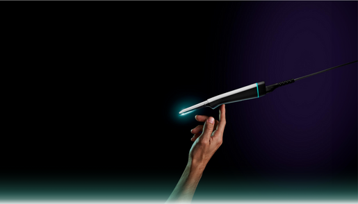
Looking for your next step?
Mastering your intraoral scanner is about far more than just owning the latest device. It’s about technique, attention to detail and a clear understanding of what the scanner can and can’t do.
Follow us on Instagram, Facebook, LinkedIn and YouTube to stay updated and understand how we can support your goals.